Intramembranous ossification
Videos
Page
Intramembranous ossification is one of the two essential processes during fetal development of the gnathostome skeletal system by which rudimentary bone tissue is created.
Intramembranous ossification is also an essential process during the natural healing of bone fractures and the rudimentary formation of bones of the head.

Transmission electron micrograph of a mesenchymal stem cell that is displaying typical ultrastructural characteristics.

Light micrograph of a nidus consisting of osteoprogenitor cells that are displaying a prominent Golgi apparatus.

Light micrograph of a nidus consisting of osteoblasts, many are displaying a prominent Golgi apparatus, that have created osteoid at its center.

Light micrograph of an undecalcified nidus consisting of rudimentary bone tissue that is lined by numerous osteoblasts.
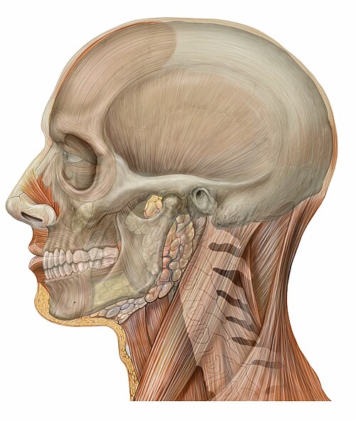
Skull
Videos
Page
The skull is a bone protective cavity for the brain. The skull is composed of four types of bone i.e., cranial bones, facial bones, ear ossicles and hyoid bone, however two parts are more prominent: the cranium and the mandible. In humans, these two parts are the neurocranium (braincase) and the viscerocranium that includes the mandible as its largest bone. The skull forms the anterior-most portion of the skeleton and is a product of cephalisation—housing the brain, and several sensory structures such as the eyes, ears, nose, and mouth. In humans, these sensory structures are part of the facial skeleton.
Skull in situ

Anatomy of a flat bone – the periosteum of the neurocranium is known as the pericranium

Chimpanzee skull

Fish head parts, 1889, Fauna of British India, Sir Francis Day