Endosomes are a collection of intracellular sorting organelles in eukaryotic cells. They are parts of endocytic membrane transport pathway originating from the trans Golgi network. Molecules or ligands internalized from the plasma membrane can follow this pathway all the way to lysosomes for degradation or can be recycled back to the cell membrane in the endocytic cycle. Molecules are also transported to endosomes from the trans Golgi network and either continue to lysosomes or recycle back to the Golgi apparatus.
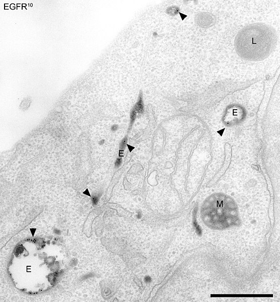
Electron micrograph of endosomes in human HeLa cells. Early endosomes (E - labeled for EGFR, 5 minutes after internalisation, and transferrin), late endosomes/MVBs (M) and lysosomes (L) are visible. Bar, 500 nm.
Diagram of the pathways that intersect endosomes in the endocytic pathway of animal cells. Examples of molecules that follow some of the pathways are shown, including receptors for EGF, transferrin, and lysosomal hydrolases. Recycling endosomes, and compartments and pathways found in more specialized cells, are not shown.
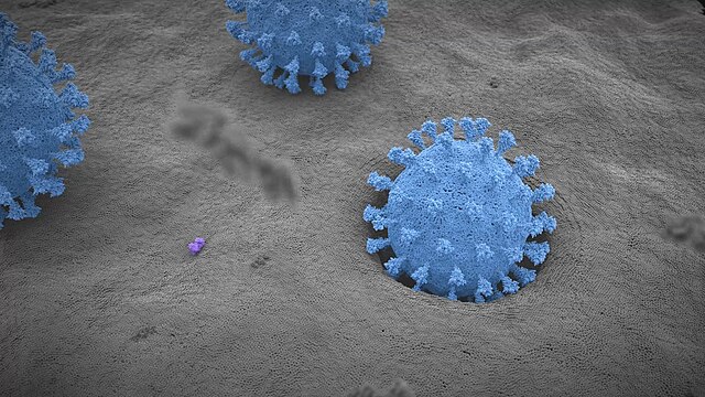
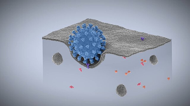
Endocytosis is a cellular process in which substances are brought into the cell. The material to be internalized is surrounded by an area of cell membrane, which then buds off inside the cell to form a vesicle containing the ingested materials. Endocytosis includes pinocytosis and phagocytosis. It is a form of active transport.
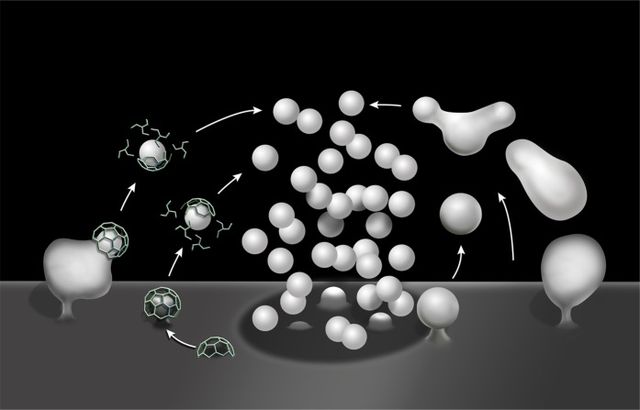
Schematic drawing illustrating clathrin-mediated (left) and clathrin-independent endocytosis (right) of synaptic vesicle membranes.
From left to right: Phagocytosis, Pinocytosis, Receptor-mediated endocytosis.
Stage 1
Stage 2


