Fetal membranes
Videos
The fetal membranes are the four extraembryonic membranes associated with the developing embryo and fetus in humans and other mammals. They are the amnion, chorion, allantois, and yolk sac. The amnion

Fetal membranes at end of second month with chorion shown in its two subdivisions

Placenta shown with attached fetal membranes

Fetal membranes illustrated at week 3 following gastrulation
Placenta
Videos
The placenta is a temporary embryonic and later fetal organ that begins developing from the blastocyst shortly after implantation. It plays critical roles in facilitating nutrient, gas, and waste exch
Human placenta from just after birth with the umbilical cord in place


Ultrasound image of human placenta and umbilical cord (color Doppler rendering) with central cord insertion and three umbilical vessels, at 20 weeks of pregnancy

Micrograph of a cytomegalovirus (CMV) infection of the placenta (CMV placentitis). The characteristic large nucleus of a CMV-infected cell is seen off-centre at the bottom-right of the image. H&E stain.
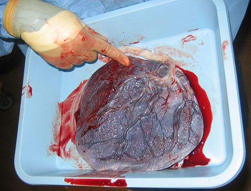
Human placenta immediately post birth.