Fibrin is a fibrous, non-globular protein involved in the clotting of blood. It is formed by the action of the protease thrombin on fibrinogen, which causes it to polymerize. The polymerized fibrin, together with platelets, forms a hemostatic plug or clot over a wound site.
Composition of a fresh thrombus at microscopy, HE stain, showing nuclear debris in a background of fibrin and red blood cells.
Micrograph showing fibrin (dark pink amorphous material) in a blocked vein surrounded by extravasated red blood cells (right of image). An artery (left of image) and the amnion (far left of image) is also seen. Placenta in a case of fetal thrombotic vasculopathy. H&E stain.
Proteins are large biomolecules and macromolecules that comprise one or more long chains of amino acid residues. Proteins perform a vast array of functions within organisms, including catalysing metabolic reactions, DNA replication, responding to stimuli, providing structure to cells and organisms, and transporting molecules from one location to another. Proteins differ from one another primarily in their sequence of amino acids, which is dictated by the nucleotide sequence of their genes, and which usually results in protein folding into a specific 3D structure that determines its activity.
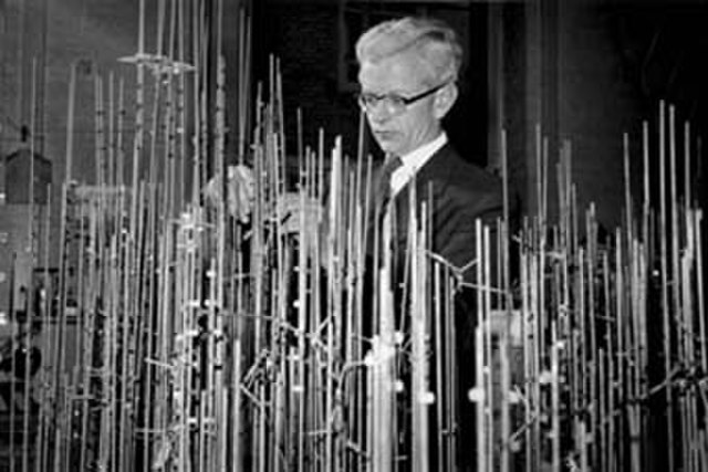
John Kendrew with model of myoglobin in progress
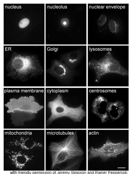
Proteins in different cellular compartments and structures tagged with green fluorescent protein (here, white)
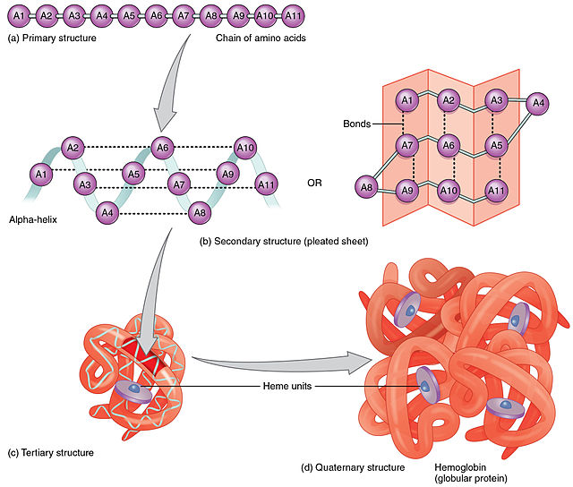
Constituent amino-acids can be analyzed to predict secondary, tertiary and quaternary protein structure, in this case hemoglobin containing heme units