Fibrinogen is a glycoprotein complex, produced in the liver, that circulates in the blood of all vertebrates. During tissue and vascular injury, it is converted enzymatically by thrombin to fibrin and then to a fibrin-based blood clot. Fibrin clots function primarily to occlude blood vessels to stop bleeding. Fibrin also binds and reduces the activity of thrombin. This activity, sometimes referred to as antithrombin I, limits clotting. Fibrin also mediates blood platelet and endothelial cell spreading, tissue fibroblast proliferation, capillary tube formation, and angiogenesis and thereby promotes revascularization and wound healing.
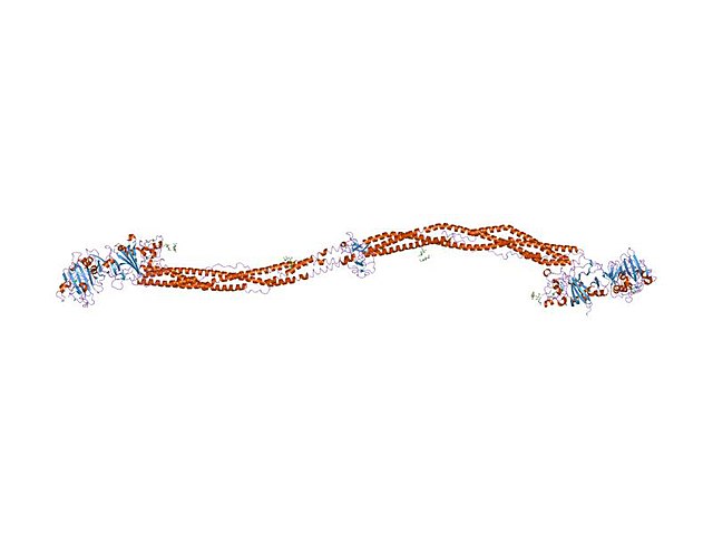
crystal structure of native chicken fibrinogen with two different bound ligands
Fibrin is a fibrous, non-globular protein involved in the clotting of blood. It is formed by the action of the protease thrombin on fibrinogen, which causes it to polymerize. The polymerized fibrin, together with platelets, forms a hemostatic plug or clot over a wound site.
Composition of a fresh thrombus at microscopy, HE stain, showing nuclear debris in a background of fibrin and red blood cells.
Micrograph showing fibrin (dark pink amorphous material) in a blocked vein surrounded by extravasated red blood cells (right of image). An artery (left of image) and the amnion (far left of image) is also seen. Placenta in a case of fetal thrombotic vasculopathy. H&E stain.