Gram stain
Videos
Page
Gram stain, is a method of staining used to classify bacterial species into two large groups: gram-positive bacteria and gram-negative bacteria. It may also be used to diagnose a fungal infection. The name comes from the Danish bacteriologist Hans Christian Gram, who developed the technique in 1884.

A Gram stain of mixed Staphylococcus aureus (S. aureus ATCC 25923, gram-positive cocci, in purple) and Escherichia coli (E. coli ATCC 11775, gram-negative bacilli, in red), the most common Gram stain reference bacteria

Gram stain of Candida albicans from a vaginal swab. The small oval chlamydospores are 2–4 µm in diameter.
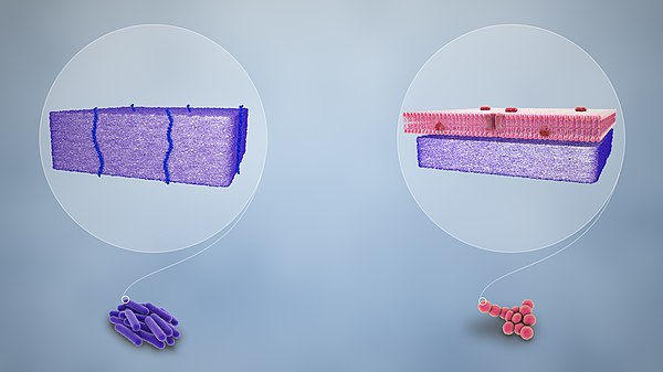
Purple-stained gram-positive (left) and pink-stained gram-negative (right)
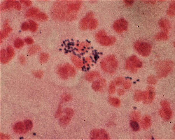
Gram-stain of gram-positive streptococci surrounded by pus cells
Staining
Videos
Page
Staining is a technique used to enhance contrast in samples, generally at the microscopic level. Stains and dyes are frequently used in histology, in cytology, and in the medical fields of histopathology, hematology, and cytopathology that focus on the study and diagnoses of diseases at the microscopic level. Stains may be used to define biological tissues, cell populations, or organelles within individual cells.

A stained histological specimen, sandwiched between a glass microscope slide.

Example of negative staining

Microscopic view of a histologic specimen of human lung tissue stained with hematoxylin and eosin.

PAS diastase showing the fungus Histoplasma.