Hematoxylin and eosin stain is one of the principal tissue stains used in histology. It is the most widely used stain in medical diagnosis and is often the gold standard. For example, when a pathologist looks at a biopsy of a suspected cancer, the histological section is likely to be stained with H&E.
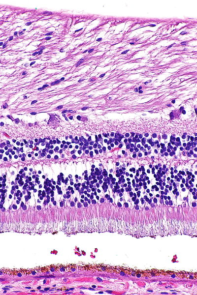
Retina (part of the eye) stained with hematoxylin and eosin, cell nuclei stained blue-purple and extracellular material stained pink.
Rack of slides being removed from a bath of hematoxylin stain.
Ductal carcinoma in situ (DCIS) in breast tissue, cell nuclei (blue-purple), extracellular material (pink).
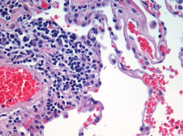
Lung tissue taken from an emphysema patient. Cell nuclei (blue-purple), red blood cells (bright red), other cell bodies and extracellular material (pink), and air spaces (white).
Staining is a technique used to enhance contrast in samples, generally at the microscopic level. Stains and dyes are frequently used in histology, in cytology, and in the medical fields of histopathology, hematology, and cytopathology that focus on the study and diagnoses of diseases at the microscopic level. Stains may be used to define biological tissues, cell populations, or organelles within individual cells.
A stained histological specimen, sandwiched between a glass microscope slide.
Example of negative staining
Microscopic view of a histologic specimen of human lung tissue stained with hematoxylin and eosin.
PAS diastase showing the fungus Histoplasma.


