Hyperpolarization (biology)
Videos
Hyperpolarization is a change in a cell's membrane potential that makes it more negative. Living cells typically have a negative resting potential. Animal excitable cells, as well as cells of other or

Diagram of membrane potential changes during an action potential

The (a) resting membrane potential is a result of different concentrations of Na+ and K+ ions inside and outside the cell. A nerve impulse causes Na+ to enter the cell, resulting in (b) depolarization. At the peak action potential, K+ channels open and the cell becomes (c) hyperpolarized.
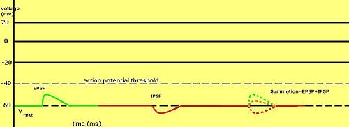
An example of inhibitory postsynaptic potentials (IPSPs), excitatory postsynaptic potentials (EPSPs), and their summation.
Depolarization
Videos
In biology, depolarization or hypopolarization is a change within a cell, during which the cell undergoes a shift in electric charge distribution, resulting in less negative charge inside the cell com
Hyperpolarization (biology)
…temporarily deviate from the resting value. This is one of many mechanisms of cell signaling. In excitable cells, activation is typically achieved through depolarization, i.e., the membrane potential deviating towards less negative values. Thus, hyperpolarization, as an opposite process, makes the cell more difficult…

Action potential in a neuron, showing depolarization, in which the cell's internal charge becomes less negative (more positive), and repolarization, where the internal charge returns to a more negative value.

Voltage-gated sodium channel. Open channel (top) carries an influx of Na+ ions, giving rise to depolarization. As the channel becomes closed/inactivated (bottom), the depolarization ends.

Structure of a neuron

Summation of stimuli at an axon hillock