Immunology
Videos
Immunology is a branch of biology and medicine that covers the study of immune systems in all organisms.
MRSA (yellow) engulfed by neutrophil (purple) Photo Source: National Institute of Allergy and Infectious Diseases


Tree of life by Haeckel
Immune system
Videos
The immune system is a network of biological systems that protects an organism from diseases. It detects and responds to a wide variety of pathogens, such as viruses, bacteria, and parasites, as well
Immunology
…Immunology is a branch of biology and medicine that covers the study of immune systems in all organisms. Immunology charts, measures, and contextualizes the physiological functioning of the immune system in states of both health and…

A scanning electron microscope image of a single neutrophil (yellow/right), engulfing anthrax bacteria (orange/left) – scale bar is 5 μm (false color)
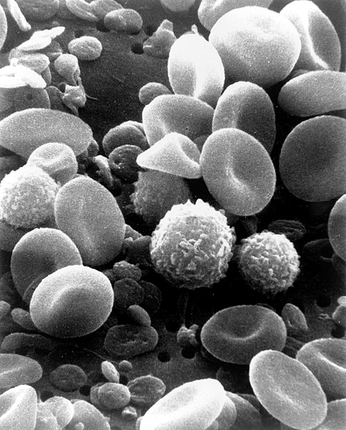
A scanning electron microscope image of normal circulating human blood. One can see red blood cells, several knobby white blood cells, including lymphocytes, a monocyte, and a neutrophil, and many small disc-shaped platelets.

An antibody is made up of two heavy chains and two light chains. The unique variable region allows an antibody to recognize its matching antigen.
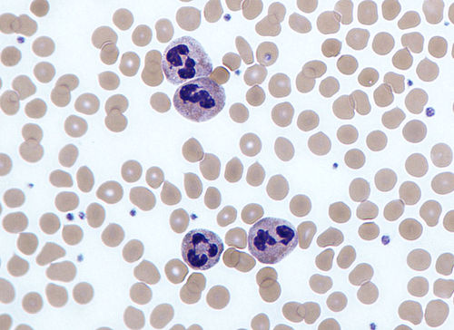
Four neutrophils in a Giemsa-stained blood film