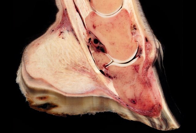
Laminitis is a disease that affects the feet of ungulates and is found mostly in horses and cattle. Clinical signs include foot tenderness progressing to inability to walk, increased digital pulses, and increased temperature in the hooves. Severe cases with outwardly visible clinical signs are known by the colloquial term founder, and progression of the disease will lead to perforation of the coffin bone through the sole of the hoof or being unable to stand up, requiring euthanasia.
Radiograph of a horse hoof showing rotation of the coffin bone and evidence of sinking, a condition often associated with laminitis. The annotation P2 stands for the middle phalanx, or pastern bone, and P3 denotes the distal phalanx, or coffin bone. The yellow lines mark the distance between the top and bottom part of the coffin bone relative to the hoof wall, showing the distal (bottom) of the coffin bone is rotated away from the hoof wall.
Hoof sagittal section with massive inflammation and rotation of third phalanx
Hoof specimen, sagittal section. Severe P3 rotation and penetration into the sole. A lamellar wedge is evident.
The front feet of this horse exhibit the rings and overgrowth typical of foundered horses.
Cattle are large, domesticated, bovid ungulates widely kept as livestock. They are prominent modern members of the subfamily Bovinae and the most widespread species of the genus Bos. Mature female cattle are called cows and mature male cattle are bulls. Young female cattle are called heifers, young male cattle are oxen or bullocks, and castrated male cattle are known as steers.
Image: Cow (Fleckvieh breed) Oeschinensee Slaunger 2009 07 07
Image: lossy page 1 GLW 2 global distributions of a) cattle.tif
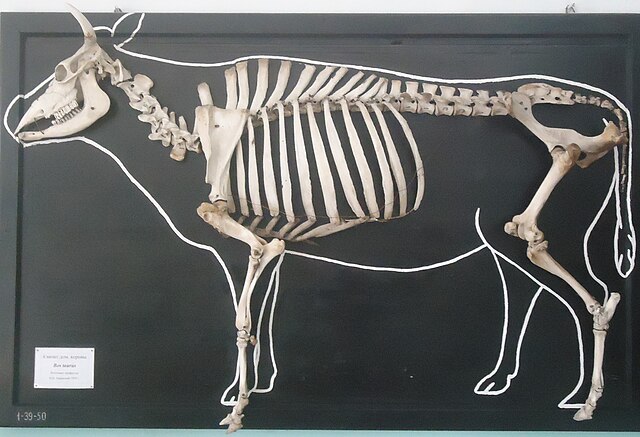
Skeleton
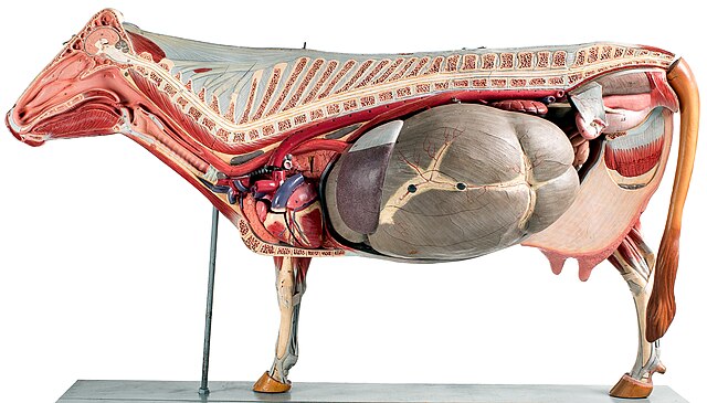
Anatomical model, showing the large 4-chambered stomach


