Lymphocyte
Videos
A lymphocyte is a type of white blood cell in the immune system of most vertebrates. Lymphocytes include T cells, B cells, and innate lymphoid cells, of which natural killer cells are an important sub

Scanning electron micrograph of a human T cell

A stained lymphocyte surrounded by red blood cells viewed using a light microscope
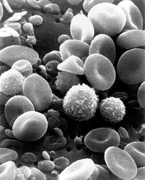
A scanning electron microscope image of normal circulating human blood showing red blood cells, several types of white blood cells including lymphocytes, a monocyte, a neutrophil and many small disc-shaped platelets

Several lymphocytes seen collected around a tuberculous granuloma
T cell
Videos
T cells are an important part of the immune system and play a central role in the adaptive immune response. T cells can be distinguished from other lymphocytes by the presence of a T-cell receptor on
Lymphocyte
…A lymphocyte is a type of white blood cell (leukocyte) in the immune system of most vertebrates. Lymphocytes include T cells (for cell-mediated and cytotoxic adaptive immunity), B cells (for humoral, antibody-driven adaptive immunity), and innate lymphoid cells (ILCs; "innate…

Scanning electron micrograph of a red blood cell (left), a platelet (center), and a T lymphocyte (right); colorized

HIV-infected T cell

Depiction of the various key subsets of CD4-positive T cells with corresponding associated cytokines and transcription factors.