Mesenchymal stem cell
Videos
Mesenchymal stem cells, also known as mesenchymal stromal cells or medicinal signaling cells, are multipotent stromal cells that can differentiate into a variety of cell types, including osteoblasts,
Transmission electron micrograph of a mesenchymal stem cell displaying typical ultrastructural characteristics


Human bone marrow-derived Mesenchymal stem cell showing fibroblast-like morphology seen under phase contrast microscope (Carl Zeiss Axiovert 40 CFL) at 63 x magnification

Typical gross appearance of a tubular cartilaginous construct engineered from amniotic mesenchymal stem cells
Osteoblast
Videos
Osteoblasts are cells with a single nucleus that synthesize bone. However, in the process of bone formation, osteoblasts function in groups of connected cells. Individual cells cannot make bone. A gro
Mesenchymal stem cell
…mesenchymal stromal cells or medicinal signaling cells, are multipotent stromal cells that can differentiate into a variety of cell types, including osteoblasts (bone-making cells), chondrocytes (cartilage cells), myocytes (muscle cells) and adipocytes (fat cells which give rise to marrow adipose tissue). The…

Osteoblasts (purple) rimming a bony spicule (pink - on diagonal of image). In this routinely fixed and decalcified (bone mineral removed) tissue, the osteoblasts have retracted and are separated from each other and from their underlying matrix. In living bone, the cells are linked by tight junctions and gap junctions, and integrated with underlying osteocytes and matrix H&E stain.

Major features of the bone-forming complex, the osteon, composed of osteoblasts and osteocytes.

Osteoblast (Wright Giemsa stain, 100x)
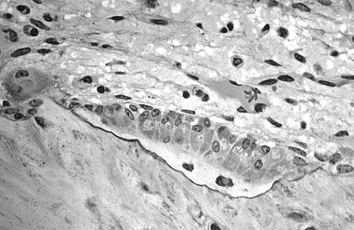
Light micrograph of decalcified cancellous bone displaying osteoblasts actively synthesizing osteoid, containing two osteocytes