Microglia
Videos
Photos
Microglia are a type of neuroglia located throughout the brain and spinal cord. Microglia account for about 10-15% of cells found within the brain. As the resident macrophage cells, they act as the first and main form of active immune defense in the central nervous system (CNS). Microglia originate in the yolk sac under a tightly regulated molecular process. These cells are distributed in large non-overlapping regions throughout the CNS. Microglia are key cells in overall brain maintenance—they are constantly scavenging the CNS for plaques, damaged or unnecessary neurons and synapses, and infectious agents. Since these processes must be efficient to prevent potentially fatal damage, microglia are extremely sensitive to even small pathological changes in the CNS. This sensitivity is achieved in part by the presence of unique potassium channels that respond to even small changes in extracellular potassium. Recent evidence shows that microglia are also key players in the sustainment of normal brain functions under healthy conditions. Microglia also constantly monitor neuronal functions through direct somatic contacts and exert neuroprotective effects when needed.

Microglia in resting state from rat cortex before traumatic brain injury (lectin staining with HRP)

Microglia/macrophage – activated form from rat cortex after traumatic brain injury (lectin staining with HRP)

Rat microglia grown in tissue culture in green, along with nerve fiber processes shown in red.

Microglia in rat cerebellar molecular layer in red, stained with antibody to IBA1/AIF1. Bergmann glia processes are shown in green, DNA in blue.
Macrophage
Videos
Photos
Macrophages are a type of white blood cell of the innate immune system that engulf and digest pathogens, such as cancer cells, microbes, cellular debris, and foreign substances, which do not have proteins that are specific to healthy body cells on their surface. This process is called phagocytosis, which acts to defend the host against infection and injury.

Gram stain of a macrophage with ingested S. epidermidis bacteria, seen as purple granules within its cytoplasm.
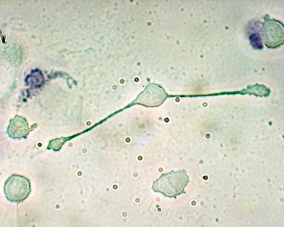
A macrophage stretching its "arms" (filopodia) to engulf two particles, possibly pathogens, in a mouse (trypan blue exclusion staining).

Siderophage

Anthracotic macrophage