Nose
Videos
A nose is a protuberance in vertebrates that houses the nostrils, or nares, which receive and expel air for respiration alongside the mouth. Behind the nose are the olfactory mucosa and the sinuses. Behind the nasal cavity, air next passes through the pharynx, shared with the digestive system, and then into the rest of the respiratory system. In humans, the nose is located centrally on the face and serves as an alternative respiratory passage especially during suckling for infants.
The protruding nose that is completely separate from the mouth part is a characteristic found only in therian mammals. It has been theorized that this unique mammalian nose evolved from the anterior part of the upper jaw of the reptilian-like ancestors (synapsids).

3D medical animation still shot depicting a human nose

The nose of a tapir

Elephants have prehensile noses.
Respiratory system
Videos
The respiratory system is a biological system consisting of specific organs and structures used for gas exchange in animals and plants. The anatomy and physiology that make this happen varies greatly, depending on the size of the organism, the environment in which it lives and its evolutionary history. In land animals, the respiratory surface is internalized as linings of the lungs. Gas exchange in the lungs occurs in millions of small air sacs; in mammals and reptiles, these are called alveoli, and in birds, they are known as atria. These microscopic air sacs have a very rich blood supply, thus bringing the air into close contact with the blood. These air sacs communicate with the external environment via a system of airways, or hollow tubes, of which the largest is the trachea, which branches in the middle of the chest into the two main bronchi. These enter the lungs where they branch into progressively narrower secondary and tertiary bronchi that branch into numerous smaller tubes, the bronchioles. In birds, the bronchioles are termed parabronchi. It is the bronchioles, or parabronchi that generally open into the microscopic alveoli in mammals and atria in birds. Air has to be pumped from the environment into the alveoli or atria by the process of breathing which involves the muscles of respiration.
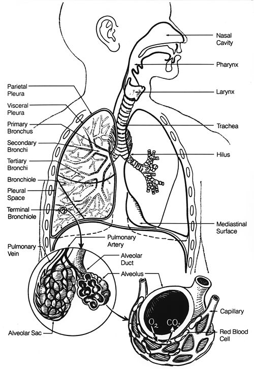

Fig. 2. The lower respiratory tract, or "Respiratory Tree" Trachea Mainstem bronchus Lobar bronchus Segmental bronchus Bronchiole Alveolar duct Alveolus

Fig. 4 The effect of the muscles of inhalation in expanding the rib cage. The particular action illustrated here is called the pump handle movement of the rib cage.

Fig. 7 The muscles of breathing at rest: inhalation on the left, exhalation on the right. Contracting muscles are shown in red; relaxed muscles in blue. Contraction of the diaphragm generally contributes the most to the expansion of the chest cavity (light blue). However, at the same time, the intercostal muscles pull the ribs upwards (their effect is indicated by arrows) also causing the rib cage to expand during inhalation (see diagram on other side of the page). The relaxation of all these muscles during exhalation causes the rib cage and abdomen (light green) to elastically return to their resting positions. Compare with Fig. 6, the MRI video of the chest movements during the breathing cycle.