The nuclear lamina is a dense fibrillar network inside the nucleus of eukaryote cells. It is composed of intermediate filaments and membrane associated proteins. Besides providing mechanical support, the nuclear lamina regulates important cellular events such as DNA replication and cell division. Additionally, it participates in chromatin organization and it anchors the nuclear pore complexes embedded in the nuclear envelope.
Confocal microscopic analysis of dermal fibroblast in primary culture from a control (a and b) and the subject with HGPS (c and d). Labelling was performed with anti-lamin A/C antibodies. Note the presence of irregularly shaped nuclear envelopes in many of the subject's fibroblasts

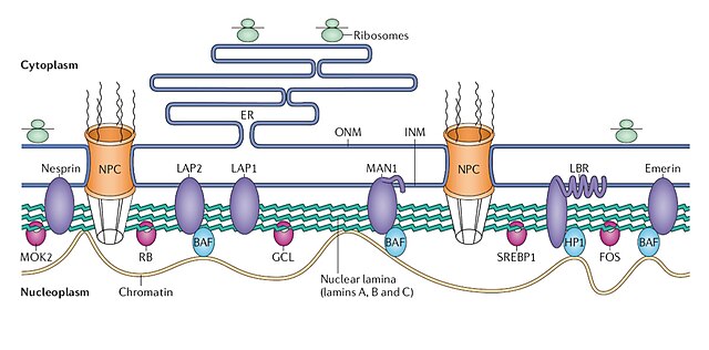
Structure and function of the nuclear lamina. The nuclear lamina lies on the inner surface of the inner nuclear membrane (INM), where it serves to maintain nuclear stability, organize chromatin and bind nuclear pore complexes (NPCs) and a steadily growing list of nuclear envelope proteins (purple) and transcription factors (pink). Nuclear envelope proteins that are bound to the lamina include nesprin, emerin, lamina-associated proteins 1 and 2 (LAP1 and LAP2), the lamin B receptor (LBR) and MAN1. Transcription factors that bind to the lamina include the retinoblastoma transcriptional regulator (RB), germ cell-less (GCL), sterol response element binding protein (SREBP1), FOS and MOK2. Barrier to autointegration factor (BAF) is a chromatin-associated protein that also binds to the nuclear lamina and several of the aforementioned nuclear envelope proteins. Heterochromatin protein 1 (HP1) binds both chromatin and the LBR. ONM, outer nuclear membrane.
The cell nucleus is a membrane-bound organelle found in eukaryotic cells. Eukaryotic cells usually have a single nucleus, but a few cell types, such as mammalian red blood cells, have no nuclei, and a few others including osteoclasts have many. The main structures making up the nucleus are the nuclear envelope, a double membrane that encloses the entire organelle and isolates its contents from the cellular cytoplasm; and the nuclear matrix, a network within the nucleus that adds mechanical support.
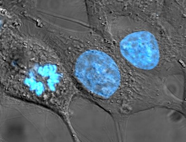
HeLa cells stained for nuclear DNA with the blue fluorescent Hoechst dye. The central and rightmost cells are in interphase, thus their entire nuclei are labeled. On the left, a cell is going through mitosis and its DNA has condensed.
An image of a newt lung cell stained with fluorescent dyes during metaphase. The mitotic spindle can be seen, stained green, attached to the two sets of chromosomes, stained light blue. All chromosomes but one are already at the metaphase plate.
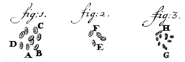
Oldest known depiction of cells and their nuclei by Antonie van Leeuwenhoek, 1719
Drawing of a Chironomus salivary gland cell published by Walther Flemming in 1882. The nucleus contains polytene chromosomes.


