Retina
Videos
The retina is the innermost, light-sensitive layer of tissue of the eye. The optics of the eye create a focused two-dimensional image of the visual world on the retina, which then processes that image

Illustration of the distribution of cone cells in the fovea of an individual with normal colour vision (left), and a colourblind (protanopic) retina. The center of the fovea holds very few blue-sensitive cones.

Macula histology (OCT)

ERP optic cabling
Central nervous system
Videos
The central nervous system is the part of the nervous system consisting primarily of the brain, and the spinal cord. The retina is also technically part of the CNS. The CNS is named so because the bra
Retina
…the optic nerve originate as outgrowths of the developing brain, specifically the embryonic diencephalon; thus, the retina is considered part of the central nervous system (CNS) and is actually brain tissue. It is the only part of the nervous system that can be visualized noninvasively. Like most of the brain, the retina…
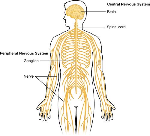
Schematic diagram showing the central and peripheral nervous system of a human

Dissection of a human brain with labels showing the clear division between white and gray matter.

Different ways in which the CNS can be activated without engaging the cortex, and making us aware of the actions. The above example shows the process in which the pupil dilates during dim light, activating neurons in the spinal cord. The second example shows the constriction of the pupil as a result of the activation of the Eddinger-Westphal nucleus (a cerebral ganglion).

Schematic image showing the locations of a few tracts of the spinal cord.