Romanowsky staining is a prototypical staining technique that was the forerunner of several distinct but similar stains widely used in hematology and cytopathology. Romanowsky-type stains are used to differentiate cells for microscopic examination in pathological specimens, especially blood and bone marrow films, and to detect parasites such as malaria within the blood.
Blood film with Giemsa stain. Monocytes surrounded by erythrocytes.
Bronchoalveolar lavage specimen stained with Diff-Quik, a commercial Romanowsky stain variant widely used in cytopathology
Dmitri Leonidovich Romanowsky (1861-1921)
Ernst Malachowsky
Staining is a technique used to enhance contrast in samples, generally at the microscopic level. Stains and dyes are frequently used in histology, in cytology, and in the medical fields of histopathology, hematology, and cytopathology that focus on the study and diagnoses of diseases at the microscopic level. Stains may be used to define biological tissues, cell populations, or organelles within individual cells.
A stained histological specimen, sandwiched between a glass microscope slide.
Example of negative staining
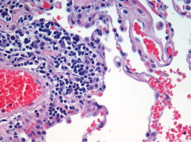
Microscopic view of a histologic specimen of human lung tissue stained with hematoxylin and eosin.
PAS diastase showing the fungus Histoplasma.


