Scanning transmission electron microscopy
A scanning transmission electron microscope (STEM) is a type of transmission electron microscope (TEM). Pronunciation is [stɛm] or [ɛsti:i:ɛm]. As with a conventional transmission electron microscope (CTEM), images are formed by electrons passing through a sufficiently thin specimen. However, unlike CTEM, in STEM the electron beam is focused to a fine spot which is then scanned over the sample in a raster illumination system constructed so that the sample is illuminated at each point with the beam parallel to the optical axis. The rastering of the beam across the sample makes STEM suitable for analytical techniques such as Z-contrast annular dark-field imaging, and spectroscopic mapping by energy dispersive X-ray (EDX) spectroscopy, or electron energy loss spectroscopy (EELS). These signals can be obtained simultaneously, allowing direct correlation of images and spectroscopic data.
An ultrahigh-vacuum STEM equipped with a 3rd-order spherical aberration corrector
Inside the aberration corrector (hexapole-hexapole type)
Schematic of a STEM with aberration corrector
Atomic resolution imaging of SrTiO3, using annular dark field (ADF) and annular bright field (ABF) detectors. Overlay: strontium (green), titanium (grey) and oxygen (red)
Transmission electron microscopy
Transmission electron microscopy (TEM) is a microscopy technique in which a beam of electrons is transmitted through a specimen to form an image. The specimen is most often an ultrathin section less than 100 nm thick or a suspension on a grid. An image is formed from the interaction of the electrons with the sample as the beam is transmitted through the specimen. The image is then magnified and focused onto an imaging device, such as a fluorescent screen, a layer of photographic film, or a detector such as a scintillator attached to a charge-coupled device or a direct electron detector.
The duplicate of an early TEM on display at the Deutsches Museum in Munich, Germany
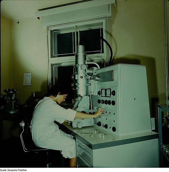
A transmission electron microscope (1976).
The electron source of the TEM is at the top, where the lensing system (4,7 and 8) focuses the beam on the specimen and then projects it onto the viewing screen (10). The beam control is on the right (13 and 14)
TEM sample support mesh "grid", with ultramicrotomy sections


