Signs and symptoms
Videos
Signs and symptoms are diagnostic indications of an illness, injury, or condition. While signs are objective and externally observable, symptoms are a person's reported subjective experiences.
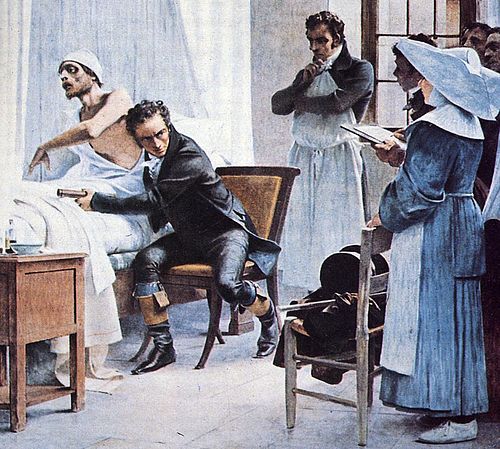
Painting of René Laennec in 1816 using an early method of auscultation on a man with tuberculosis
Immune system
Videos
The immune system is a network of biological systems that protects an organism from diseases. It detects and responds to a wide variety of pathogens, such as viruses, bacteria, and parasites, as well
Signs and symptoms
…of the ordinary that is experienced by an individual such as feeling feverish, a headache or other pains in the body. Symptoms can be a result of the immune system's response to an infection, the physical manifestation of an abnormal body condition, or the effect of a consumed substance. Symptoms, though often…

A scanning electron microscope image of a single neutrophil (yellow/right), engulfing anthrax bacteria (orange/left) – scale bar is 5 μm (false color)
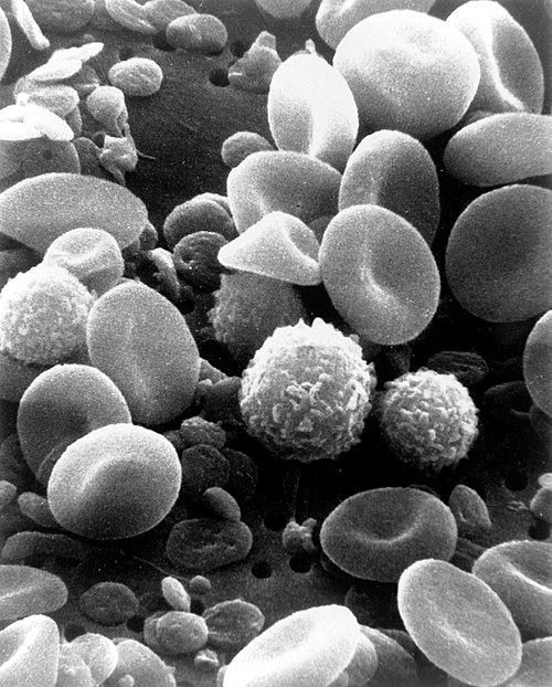
A scanning electron microscope image of normal circulating human blood. One can see red blood cells, several knobby white blood cells, including lymphocytes, a monocyte, and a neutrophil, and many small disc-shaped platelets.

An antibody is made up of two heavy chains and two light chains. The unique variable region allows an antibody to recognize its matching antigen.
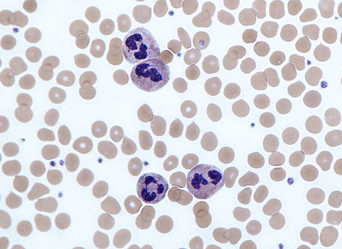
Four neutrophils in a Giemsa-stained blood film