Striated muscle tissue
Videos
Striated muscle tissue is a muscle tissue that features repeating functional units called sarcomeres. The presence of sarcomeres manifests as a series of bands visible along the muscle fibers, which is responsible for the striated appearance observed in microscopic images of this tissue. There are two types of striated muscle:Cardiac muscle
Skeletal muscle

Micrograph of HPS stained skeletal striated muscle (fibularis longus).
Skeletal muscle
Videos
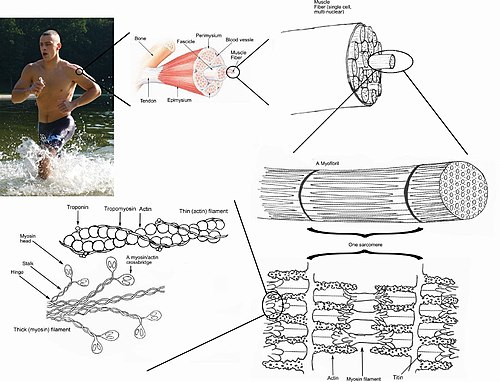
Skeletal muscles are organs of the vertebrate muscular system and typically are attached by tendons to bones of a skeleton. The muscle cells of skeletal muscles are much longer than in the other types of muscle tissue, and are often known as muscle fibers. The muscle tissue of a skeletal muscle is striated – having a striped appearance due to the arrangement of the sarcomeres.
A top-down view of skeletal muscle

ATPase staining of a muscle cross section. Type II fibers are dark, due to the alkaline pH of the preparation. In this example, the size of the type II fibers is considerably less than the type I fibers due to denervation atrophy.

Diagram of sarcoplasmic reticulum with terminal cisternae and T-tubules.

When a sarcomere contracts, the Z lines move closer together, and the I band becomes smaller. The A band stays the same width. At full contraction, the thin and thick filaments overlap.