Vasoactive intestinal peptide
Vasoactive intestinal peptide, also known as vasoactive intestinal polypeptide or VIP, is a peptide hormone that is vasoactive in the intestine. VIP is a peptide of 28 amino acid residues that belongs to a glucagon/secretin superfamily, the ligand of class II G protein–coupled receptors.
VIP is produced in many tissues of vertebrates including the gut, pancreas, cortex, and suprachiasmatic nuclei of the hypothalamus in the brain. VIP stimulates contractility in the heart, causes vasodilation, increases glycogenolysis, lowers arterial blood pressure and relaxes the smooth muscle of trachea, stomach and gallbladder. In humans, the vasoactive intestinal peptide is encoded by the VIP gene.
Suprachiasmatic nucleus is shown in green.
G protein-coupled receptor
G protein-coupled receptors (GPCRs), also known as seven-(pass)-transmembrane domain receptors, 7TM receptors, heptahelical receptors, serpentine receptors, and G protein-linked receptors (GPLR), form a large group of evolutionarily related proteins that are cell surface receptors that detect molecules outside the cell and activate cellular responses. They are coupled with G proteins. They pass through the cell membrane seven times in the form of six loops of amino acid residues, which is why they are sometimes referred to as seven-transmembrane receptors. Ligands can bind either to the extracellular N-terminus and loops or to the binding site within transmembrane helices. They are all activated by agonists, although a spontaneous auto-activation of an empty receptor has also been observed.

Cartoon depicting the basic concept of GPCR conformational activation. Ligand binding disrupts an ionic lock between the E/DRY motif of TM-3 and acidic residues of TM-6. As a result, the GPCR reorganizes to allow activation of G-alpha proteins. The "side perspective" is a view from above and to the side of the GPCR as it is set in the plasma membrane (the membrane lipids have been omitted for clarity). The incorrectly labelled "intracellular perspective" shows an extracellular view looking down at the plasma membrane from outside the cell.
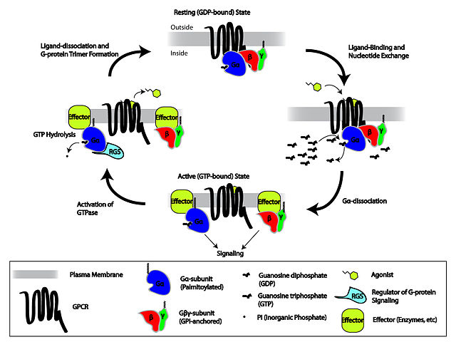
Cartoon depicting the heterotrimeric G-protein activation/deactivation cycle in the context of GPCR signaling
The effect of Rs and Gs in cAMP signal pathway
The effect of Ri and Gi in cAMP signal pathway


