Ventral tegmental area
Videos
The ventral tegmental area, also known as the ventral tegmental area of Tsai, or simply ventral tegmentum, is a group of neurons located close to the midline on the floor of the midbrain. The VTA is t

Ventral tegmentum shown in the tegmentum
Locus coeruleus
Videos
The locus coeruleus, also spelled locus caeruleus or locus ceruleus, is a nucleus in the pons of the brainstem involved with physiological responses to stress and panic. It is a part of the reticular
Micrograph showing the locus coeruleus (upper-right of image) in an axial section of the pons. The fourth ventricle (quasi-triangular white area) is in the upper-left of the image. The midline is seen on the left. The large white area in the upper-left corner is where the cerebellum would be. HE-LFB stain.


Locus coeruleus highlighted in green.
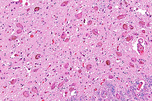
Micrograph showing the locus coeruleus. HE-LFB stain