Zymography is an electrophoretic technique for the detection of hydrolytic enzymes, based on the substrate repertoire of the enzyme. Three types of zymography are used; in gel zymography, in situ zymography and in vivo zymography. For instance, gelatin embedded in a polyacrylamide gel will be digested by active gelatinases run through the gel. After Coomassie staining, areas of degradation are visible as clear bands against a darkly stained background.
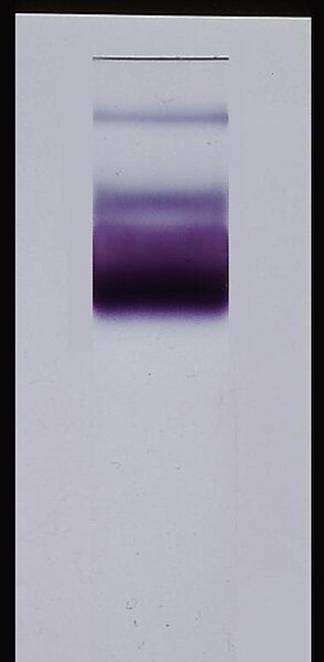
Hemoglobinase imprint of Plasmodium knowlesi.
Gel electrophoresis is a method for separation and analysis of biomacromolecules and their fragments, based on their size and charge. It is used in clinical chemistry to separate proteins by charge or size and in biochemistry and molecular biology to separate a mixed population of DNA and RNA fragments by length, to estimate the size of DNA and RNA fragments or to separate proteins by charge.
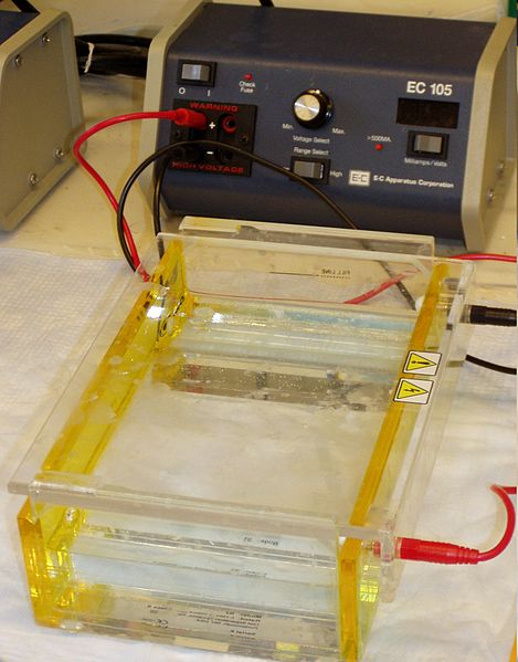
Gel electrophoresis apparatus – an agarose gel is placed in this buffer-filled box and an electric current is applied via the power supply to the rear. The negative terminal is at the far end (black wire), so DNA migrates toward the positively charged anode(red wire). This occurs because phosphate groups found in the DNA fragments possess a negative charge which is repelled by the negatively charged cathode and are attracted to the positively charged anode.
Inserting the gel comb in an agarose gel electrophoresis chamber
Specific enzyme-linked staining: Glucose-6-Phosphate Dehydrogenase isoenzymes in Plasmodium falciparum infected Red blood cells


